メニュー

Mohs surgery for basal cell carcinoma / squamous cell carcinoma

中文説明Click


For medical doctors, health care workers, and medical personnel

When referring your patients, we request you to kindly lend the glass slide of the biopsy as well as the pathology report. We will return the glass slide upon completing the surgery. The case report from us is the first Mohs micrographic surgery paper from Japan, which is published from a major international dermatology journal: Suzuki et al. Barriers to Mohs Micrographic Surgery in Japanese Patients With Basal Cell Carcinoma. Cutis. 2024 Jul; 114(1): E16-20. doi; 10.12788/cutis.1057. 


For the patients who are considering taking Mohs surgery

An appointment is necessary for a consultation on Mohs surgery. Mohs surgery is not covered by Japanese Health Insurance. If you would like to undergo Mohs surgery, please provide us the following: a) Referral letter from the previous doctor, b) The pathology report of the biopsy, and c) The glass slide or the biopsy.

Before you start browsing this site, please be aware that the information below includes some surgical images. Unauthorized reproduction of these images is prohibited by law.


What is Mohs surgery?

It is formally termed as Mohs micrographic surgery, which is named after Dr. Frederic Mohs who started this technique on June 30, 1936. It is a surgical technique to examine the entire cut surface of the specimen under immediately after the excision. Due to the high cure rate and sparing of normal tissue, this technique has become the gold standard treatment for facial and special-site nonmelanoma skin cancer worldwide. The five-year cure rate of basal cell carcinoma, for example, is 99% with Mohs surgery, substantially higher than the 90% of conventional wide excisions (See the table at the bottom of this site). Our clinic is the first in Japan which reported Mohs surgery cases, and is the only facility to perform Mohs surgery in the entire country as of September 2025. S Suzuki, SI Kim, TK Barlow. Barriers to Mohs Micrographic Surgery in Japanese Patients With Basal Cell Carcinoma. Cutis. 2024; 114(7): E16-20. doi: 10.12788/cutis.1057.


Mohs surgery and conventional wide excision

Conventional wide local excision is a commonly practiced technique in Japan for skin cancer removal. It takes 3-10 mm margin of normal skin from the grossly visible tumor edge. The majority of the skin within this 3-10 mm range is normal skin with no need for removal (The red line in Figure 1 is an example of the excision line with 4 mm margin). Figure 2 shows additional spindle cut line to be able to close the skin defect with suture. In this example, the skin cancer illustrated in brown color may be left underneath. The specimen will be sent to the outside laboratory. At the laboratory, the specimen is fixed with formalin and then vertically sectioned every 2 to 3 mm (bread-loafed; Fig. 3) to create a thin representative slice. Each representative slice appears to show clear margins. In reality, the tumor remains between the second and the third slices, which leads to a false-positive interpretation. This entire process of pathological examination takes 10-14 days. Because of the representative nature of this method, the long-term prognosis (such as 5-year / 10-year recurrence rate) is worse than for Mohs surgery. Even when positive margins are identified on pathology, the lack of precision on the exact location of the residual tumor will require the surgeon to excise the entire scar, resulting in an unnecessary large surgical defect. This secondary excision also requires another 10-14 days before receiving the pathology report. Until the surgeon confirms that no skin cancer cell is left, the skin defect after excision may be left open, or the skin cancer cells may remain if closed before the conclusion of the pathology report.


With Mohs surgery, the tumor is excised with a 1-2 mm margin of normal skin, leaving most of the surrounding normal skin intact (Fig. 4). There are small incisions at the 12-, 3-, 6-, and 9-o’clock positions to provide orientation (Fig. 5). The specimen’s entire cut surface is placed en face on a plane, frozen, cut and mounted on a glass slide (Fig. 6). Unlike the conventional wide local excision, the entire cut surface is examined. It is stained with hematoxylin and eosin and evaluated by the Mohs surgeon, who examines the glass slide under a microscope to determine the presence of tumor cells and draws a map of any residual tumor location(s) (Fig.7). In this example, tumor cells are seen as dark brown around the 4-o’clock position in the superficial to mid dermis. If the residual tumor is identified, an additional stage of excision restricted to the precise site of tumor cells is performed, and the similar specimen analysis is undertaken until the surgeon confirms that no residual tumor cells remain. Once no residual tumor cells are identified, immediate repair of the skin defect is performed on the same day. The entire process of the pathological evaluation of the specimen takes 1-2 hours. Mohs surgery would enable to sparing of normal skin as much as possible, and also the prognosis, such as the 5-year recurrence rate, is better than with the conventional wide local excision.


Recent trends of skin cancer (basal cell carcinoma and squamous cell carcinoma) and Mohs surgery

Due to the high cure rate and sparing of normal tissue, Mohs surgery has become the gold standard treatment for facial and special-site nonmelanoma skin cancer worldwide. Mohs surgery is performed on more than 876,000 tumors annually in the United States.1 Among the 3.5 million Americans diagnosed with nonmelanoma skin cancer in 2006, one-quarter were treated with Mohs surgery.2 In Japan, basal cell carcinoma is the most common skin cancer, with an incidence of 3.34 cases per 100,000 individuals; squamous cell carcinoma is the second most common, with an incidence of 2.5 cases per 100,000 individuals. The guideline of skin cancer by Japanese Dermatological Association indicates Mohs surgery as an effective surgical method, but as of September 2025, no other clinics/hospitals in Japan perform Mohs surgery, and the majority of cases are treated with the conventional wide local excision.4 Japanese National Health Insurance does not cover Mohs surgery.


The actual Mohs surgery procedure

Mohs surgery is a day-surgery under local anesthesia.


a) Incision line with 1-2 mm margin is designed (Fig.4, above). Notches of 4 corners are added to ensure the orientation.


b) Tumor is excised, a gauze with saline is applied, and wait for 1-2 hours while examining the specimen. The patient can eat or walk around until the histological result is ready. The specimen is dyed with color, marked with nylon suture for orientation, then the cut surface is placed en-face and frozen (Fig. 8 and 9).


c) The specimen is sliced with a device called a cryostat, which freezes the specimen down to -30 degree Celsius and cut with a thin slice (movie 1). The sliced specimens are mounted on glass slides and stained with the autostainer (movie 2, Fig. 10).

Movie 1 The specimen is sliced with cryostat.

Moive 2 The sliced specimens are mounted on glass slides, and are stained with the autostainer.


d) The glass slides are observed under a microscope to determine whether any residual tumor cells are exposed at the cut surface (Fig. 11). If there are any, the precise location in the specimen is located to specify the area necessary for additional excision (Fig. 12-15).


e) The result of the microscopic examination is explained to the patient. If there are any residual tumor cells, only that specific area is excised subsequently, and the same process of a)-d) above is repeated. In this case, an additional excision at 3 and 10 o’clock directions is made (Fig. 16). Just as in the first stage, a microscopic examination is conducted (Fig. 17). In this case, there are no residual tumor cells on the cut surface from the second stage excision specimens (Fig. 18 and 19). One report says that the average number of stages is 1.74.5


f) After confirming that no residual tumor cells are left, the skin defect will be reconstructed. Simple closure with suture is made, if the skin defect is small enough (Figs. 20-22). As is mentioned in the latter part, Mohs surgery is best indicated in the area with little excessive skin, such as the face, head & neck, shin, genital area, hand & foot, ankle, areola, and nipple. Depending on the location and the size of the final skin defect, a skin flap and skin grafting might be necessary to reconstruct the defect.


Comparison of the reconstruction between conventional wide local excision and Mohs surgery

Surgery and reconstruction process of 2 cases of basal cell carcinoma, infiltrative, on the similar location (nose tip) was compared between a) Mohs surgery and b) conventional wide local excision.


a) A case with Mohs surgery

This case is a basal cell carcinoma, infiltrative subtype on the nose tip. This subtype of basal cell carcinoma has a higher recurrence rate than other basal cell carcinomas. Figure 23 shows a red spot on the left side of the nose tip, which was diagnosed as basal cell carcinoma after a biopsy at the previous clinic. Under the microscope, tumor cells are seen forming clusters of spiky shape in the dermis, surrounded by strong fibrosis, exhibiting the features of infiltrative subtype of basal cell carcinoma (Fig. 24). If this subtype of basal cell carcinoma is treated surgically with conventional wide local excision, at least a 5 mm margin would be suggested, which would cause a major nose deformity. The excision line of Mohs surgery with a 1.5mm margin and notches of 4 corners are designed (Fig. 25). The excision was done under local anesthesia and an additional marking suture was put at the 12 o’clock direction to ensure that any residual tumor cells are located precisely (Fig. 26). Fortunately, this case turned out to be free of residual tumor cells after the 1st stage, and the reconstruction of the skin defect was conducted 2 hours after the excision. Because the skin defect after Mohs surgery is very small, a local flap called “rhomboid flap” was sufficient to cover the defect (Fig. 27). The flap was raised under local anesthesia and the defect was covered with the flap (Figs. 28-30). Seven months later, the scar is insignificant and nose deformity is not observed (Fig. 31). Thus, Mohs surgery completes the entire process of excision of the tumor to reconstruction under local anesthesia within the same day. Surrounding normal skin is preserved as much as possible, which makes the reconstruction of the skin defect easier than conventional wide local excision.


b) A case with conventional wide local excision

This case is also basal cell carcinoma, infiltrative type, on the nose tip (Fig. 32). The part with erosion (red part) is a recurrent tumor from the previous conventional wide local excision. Considering the high likelihood of recurrence with conventinal wide local exicison again, a 1cm margin was taken for this case (Fig. 33). The entire right side of the nose, including cartilage, was lost with the surgery, and a skin flap from the forehead and the right cheek, as well as a cartilage graft from the ear were designed to reconstruct the nose defect under general anesthesia (Figs. 34-36). The flap from the forehead can be cut if given enough time for neovascularization from the nose to be established, and it was cut one month after the flap formation surgery, again under general anesthesia (Figs. 37-39). In this example, the patient needed to stay in the hospital for over one month, and 2 surgeries under general anesthesia were required, with finally the skin defect able to be reconstructed, although the nose deformity was inevitable due to the large size skin defect (Fig. 40). Even after such an extensive removal of skin, there is higher recurrence rate than Mohs surgery, especially after the removal of a recurrent basal cell carcinoma.


Examples of good indication for Mohs surgery of basal cell carcinoma and squamous cell carcinoma

Size: larger than 5 mm

Location: Face, head and neck, shin, genital area, hand and foot, ankle, areola, and nipple

Histological subtypes: 
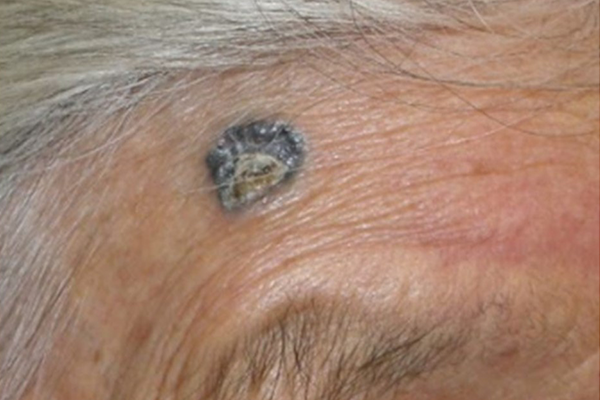
Basal cell carcinoma with the following features; Morpheaform/fibrosing/sclerosing/infiltrating/perineural/metatypical/keratotic/micronodular

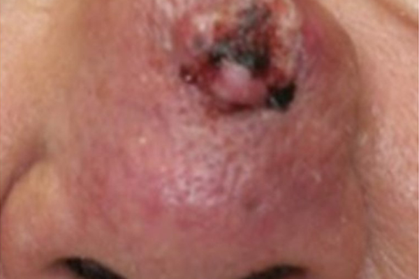
Squamous cell carcinoma with the following features;

Sclerosing/basosquamous/small cell/ poorly or undifferentiated/perineural/perivascular/ spindle cell/ pagetoid/infiltrating/keratoacanthoma type/single cell/clear cell/lymphoepithelial/sarcomatoid/Breslow depth greater than 2mm/Clark lever IV or deeper


Prognosis after Mohs surgery

The recurrence rate after Mohs surgery is lower than for conventional wide local excisions, both for basal cell carcinoma and squamous cell carcinoma, and is reported to be 1-5 % depending on the histological subtypes and observation period. One study indicates a recurrence rate of primary basal cell carcinoma of high risk subtypes 10 years following surgery of 4.4% with Mohs surgery versus 12.2% with a conventional wide local exicison.6


Surgical statistics at our clinic

As of September 2025, our clinic has treated over 140 cases with Mohs surgery, which is the only Mohs surgery facility in Japan. A case report from our clinic is published in one of the major international dermatology journals, which is the first case report of Mohs surgery in Japan.7


Fees (tax included / deductible if filing a tax return in Japan / not covered by Japanese Health Insurance)

First stage: 550,000 yen

Each additional stage: 275,000 yen

The average number of stages to be free from tumor cells: 1-3 stages (1.74 over all5)

If a local flap or skin grafting is necessary / the lesion is too large to be processed as a single specimen, an additional charge is required.


References

  1. Asgari MM, Olson J, Alam M. Needs assessment for Mohs micrographic surgery. Dermatol Clin. 2012; 30: 167-75. doi: 1016/j.det.2011.08.010
  2. Connolly SM, Baker DR, et al. AAD/ACMS/ASDSA/ASMS 2012 appropriate use criteria for Mohs micrographic surgery: a report of the American Academy of Dermatology, American College of Mohs Surgery, American Society for Dermatologic Surgery Association, and the American Society for Mohs Surgery. J Am Acad Dermatol. 2012; 67: 531-50.
  3. Ansai SI, Umebahashi Y, Katsunuma N, et al. Japanese Dermatological Association Guidelines: outlines of guidelines for cutaneous squamous cell carcinoma 2020. J Dermatol. 2021; 48: E288-311.
  4. Ad Hoc Task Force1 Connolly SM, Baker DR. AAD/ACMS/ASDSA/ASMS 2012 appropriate use criteria for Mohs micrographic surgery. J Am Acad Dermatol. 2012; 67(4):531-50. 
  5. Kristian A, Xu T, Hutfless S, et al. Outlier Practice Patterns in Mohs Micrographic Surgery: Defining the Problem and a Proposed Solution. JAMA Dermatol. 2017; 153(6): 565-70.
  6. Van Loo, Mosterd K, Krekels GA. Surgical excision versus Mohs' micrographic surgery for basal cell carcinoma of the face. Eur J Cancer. 2014; 50(17):3011-20.
  7. Suzuki S, Barriers to Mohs Micrographic Surgery in Japanese Patients With Basal Cell Carcinoma. Cutis. 2024; 114(1): E16-20.

HOME

▲ ページのトップに戻る

Close

HOME